The Wisdom Tooth We Didn’t Take Out
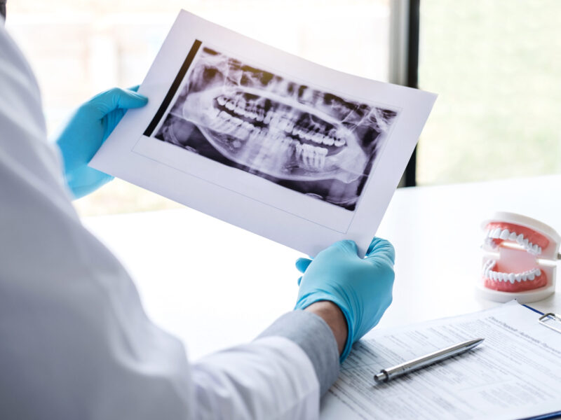
On a panoramic X-ray, a lower wisdom tooth can look like a simple job. A clear enough angle, roots that seem to sit clear of trouble, a fee, and a date in the book. That was nearly the plan for one of our patients earlier this year: a woman in her thirties, booked to have a lower wisdom tooth taken out here in the chair. Then our Brisbane dentists looked at her wisdom tooth 3D scan. The roots sat closer to the nerve than a flat picture can ever really show, and there was a shadow that we suspect might have been a cyst. So we didn’t perform wisdom tooth removal that day. What changed our treatment plan for her wasn’t the wisdom tooth itself. It was the difference between a flat X-ray and a CBCT scan that shows depth.
What a Panoramic (OPG) X-ray Shows and Where It Stops
A panoramic X-ray, or OPG, is the single wide image that captures your whole mouth in one pass. For most wisdom teeth, it tells us everything we need to plan the removal. For a few, it raises a question it can’t quite answer on its own.
What a Panoramic (OPG) X-ray Shows Clearly
- The whole mouth at once: All of your teeth at once, top and bottom, including wisdom teeth still sitting under the gum, plus the jawbone, the sinuses, and the jaw joints, in one picture.
- For a wisdom tooth: how many there are, which way each one is pointing, how deep it sits, and the rough shape and number of its roots.
- Obvious problems: Visible problems around the tooth, such as decay, bone loss, or a cyst large enough to show up.
- Low dose, used first: It does all this at a low radiation dose, taken only when there’s a clear reason to image, which is why an OPG is the usual first image before a removal.
What it can only hint at
- It’s a flat image: An OPG is flat. It presses a three-dimensional jaw into a two-dimensional picture, so structures that are genuinely apart can look like they sit on top of each other.
- Roots near the nerve: The lower wisdom tooth roots and the nerve canal (the channel that carries feeling to your lower lip and chin) often look like they meet on an OPG. The image can flag warning signs that they might be close: a darkening where the roots cross the canal, the bright lines of the canal looking broken or pushed off course, or roots that look narrowed or bent.
- A hint, not proof: These signs are a reason to look closer, not proof of contact. In one surgical study, an OPG that suggested the tooth and nerve were touching was confirmed only about one time in eight.

What CBCT (a 3D scan) Adds
If the OPG leaves a real question about the nerve, the next step is a CBCT, a scan that builds the same area in three dimensions. It doesn’t replace the OPG. It answers the one thing a flat image can’t, and for a small number of people, what it shows changes our plan.
What Becomes Visible
- The nerve in three dimensions: A CBCT shows us whether the nerve canal runs to the cheek side of the roots, the tongue side, or directly between them, the exact thing a flat OPG can’t place.
- Whether bone still separates them: It shows if there’s a thin layer of bone between the patient’s tooth and the nerve, or none at all. Direct contact, with no bone in between, is the finding that matters most for risk.
- The real shape of the roots: Extra roots, hooks, or roots that curl around the canal show up clearly, where a flat image can blur them together.
- The full size of a lesion: If the OPG hinted at something like a cyst, the scan shows its true size and spread in three dimensions.
When a 3D Scan Changes the Plan
- It can switch the approach: In one study of these closer cases, the 3D view changed the surgical plan for about one in eight, most often by moving to a nerve-sparing option rather than a straight removal.
- A nerve-sparing option: When the tooth sits hard against the nerve, the plan may shift to a coronectomy, where the crown is taken off, and the root tips are deliberately left in place to protect the nerve, or to a referral to a specialist.
- A clearer consent conversation: Even when the plan doesn’t change, the scan sharpens what we can tell you about your own risk before you decide.
She was one of them. Her 3D scan confirmed how close those roots really sat to the nerve, with what looked like it might be a cyst beside them.

What a Panoramic (OPG) X-ray Can’t Show at All
- Depth and direction: The true position in three dimensions: whether the nerve runs to the cheek side of the roots, the tongue side, or straight between them. A flat image can’t place it in that third direction.
- The bone around the nerve: Whether the thin wall of bone around the nerve is still intact or already worn through, which is part of what changes the risk.
- The true size of a lesion: The full size and shape of a lesion, like a suspected cyst, in three dimensions.
For most people, the OPG answers the question, and the tooth comes out as planned. When those warning signs appear, that is when a 3D scan earns its place.

The Signs on an OPG That Make Us Reach for a CBCT
We don’t order a 3D scan for every wisdom tooth. Most don’t need one. We order a CBCT when the flat X-ray shows specific signs that the tooth and nerve may be close together. These are the recognised warning signs we look for.
The Warning Signs We Look For
- A dark band across the roots: Where the roots cross the nerve canal, they can look darker, like a shadow banded over them, a sign that the root and the canal may be in close contact.
- The canal’s bright lines break: The nerve canal shows as two thin white lines on an OPG. Where those lines look broken or faded as a root crosses, the tooth and canal may be touching. This is one of the most reliable signs.
- The canal changes course: The canal usually runs in a smooth line. Where it bends or detours around a root, that change of course is another of the strongest signs.
- The canal narrows: The canal looks pinched where it meets the root, thinner there than on either side of it.
- The roots pinch, hook, or split: Roots that look narrowed, bent, or split into a dark fork right where they reach the canal.
How We Weigh Them
- One can be enough: A single clear sign is reason enough to stop and look closer. We don’t wait for all of them to line up.
- We read the whole picture: No sign is read on its own. We weigh it against the angle of the tooth, your age, the rest of the X-ray, and what we can see and feel in your mouth.
- Only if it changes the plan: Spotting a sign isn’t an automatic scan. We order a CBCT when the answer would actually change how, or whether, we take the tooth out, in line with the principle that imaging is taken only when there’s a clear benefit.

Why the Nerve’s Position Matters
Lower wisdom teeth grow close to a nerve that gives feeling to your lower lip, chin, and tongue. When a tooth sits right next to it, taking it out carries a small risk of bruising or stretching that nerve, which can leave the area feeling numb or tingly for a while. In most cases, that feeling fades on its own as the nerve recovers, often within a couple of months. Lasting change is rare: in a large Australian study of more than 11,000 lower wisdom teeth, it affected well under 1 in 100. Rare, but not zero, which is the honest way to put it. This is exactly why we read the X-ray carefully and look in 3D when the signs are there, so we can plan around the nerve rather than meet a surprise partway through. We don’t mean to alarm you. Most people have their wisdom teeth out with no lasting effects, and the care we take beforehand is how we keep it that way.

Medical Disclaimer
This article is general information only. It explains how wisdom tooth X-rays and 3D scans are used, but it isn’t dental or medical advice, and it can’t tell you what’s right for your own teeth. Every mouth is different, and the only way to know what suits your situation is a proper assessment by a registered dental practitioner who can examine you and look at your own images.

Frequently Asked Questions
Does everyone need a 3D scan for wisdom teeth?
No. Most wisdom teeth are planned from a single panoramic X-ray, and that is all they need. A 3D scan is only worth taking when the flat image suggests the roots may be sitting close to the nerve, or when there’s something like a possible cyst to look into. If your dentist doesn’t suggest one, that usually means the OPG has already answered the question without it.
Is a CBCT more radiation than an OPG?
Yes, a 3D scan uses more than a panoramic, but both are low. A panoramic X-ray is roughly two to three days’ worth of the natural background radiation we all get from the environment anyway. A focused 3D scan of a single area is more than that, often somewhere between several days and a couple of weeks of background radiation, and still well below a hospital medical CT scan. That balance is the reason a CBCT is taken only when it will change the plan, not as a routine extra.
Why might I be referred to a specialist?
If a lower wisdom tooth sits very close to the nerve, or there’s a lesion to deal with, the safest place to have it managed can be with an oral and maxillofacial surgeon, who does this kind of complex removal day in, day out. A referral isn’t a sign that something has gone wrong. It usually means your dentist has read the warning signs early and wants the tooth handled in the right setting, sometimes with a nerve-sparing option on the table.

Does Your Wisdom Tooth Need a Closer Look?
If a wisdom tooth has been on your mind, the simplest next step is to have it looked at properly before anything is decided. Call us on 07 3343 4869 to book an assessment, where you’ll see your own X-ray on the screen and hear exactly what it shows.
For treatment that genuinely can’t wait, we can explain your options, from Humm payment plans to early release of super through SuperCare for those who qualify.